


 14 / 22

© 2016, BSM Consulting
12
Basics of Glaucoma
Scanning Computerized Diagnostic Imaging (SCODI):
Confocal scanning laser ophthalmoscopy
creates a three-dimensional image of the optic nerve head. The optical design of instruments using
confocal scanning laser technology allows for a series of tomographic ‘‘slices,” or optical sections, of the
structure. The images acquired by this method are stored as a computer data file and manipulated to
reconstruct the three-dimensional structure, display the image, and perform data analysis. Parameters
such as cup area, cup volume, rim volume, cup-disc ratio, and peripapillary nerve fiber layer thickness are
then calculated.
Techniques such as scanning laser polarimetry and optical coherence tomography have been used to
acquire images of the retinal nerve fiber layer. The scanning laser polarimeter is essentially a scanning
laser ophthalmoscope outfitted with a polarization modulator and detector to take advantage of the
refractive properties of the retinal nerve fiber layer.
Optical coherence tomography (OCT) uses interferometry and low coherence light to obtain a high-
resolution cross section of biological structures. The Heidelberg Retinal Tomograph (HRT) employs a
confocal scanning laser ophthalmoscope to capture its cross sections of the optic nerve fiber layer. This
testing allows for comparison of the optic nerve from one testing session to another and provides
important information regarding the possible progression of optic nerve damage.
Gonioscopy
Gonioscopy is the evaluation of the eye's angle structure usually using a specialized, mirrored contact
lens while the patient is seated at the slit lamp. Common lenses are the Koeppe lens or the three- or four-
mirror lenses. The mirror is necessary to see into the angle between the iris and the cornea. Not only
does gonioscopy provide the physician with a three-dimensional view of the anterior chamber angle, it
can also be helpful in assessing the depth of the angle structure and in differentiating between narrow
angles and angle closure glaucoma. The depth of the angle is usually noted, often along with the
pigmentation within the trabecular meshwork, presence of adhesions (peripheral anterior synechia), and
presence or absence of new blood vessel growth. Compression (indentation) gonioscopy can determine if
an anatomically narrow angle is likely to be openable with iridotomy or if it is permanently closed due to
adhesions (chronic angle closure).
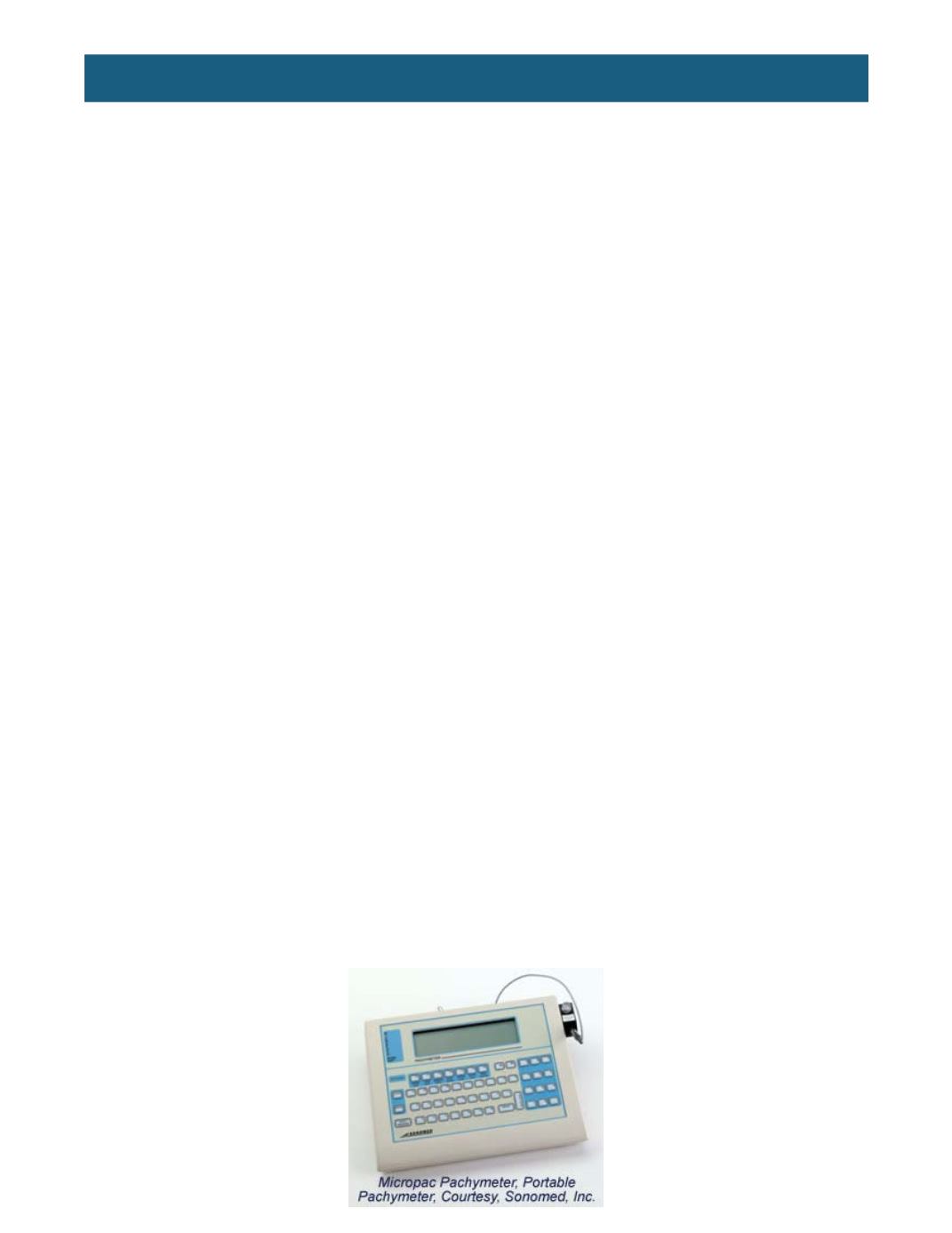
Pachymetry
The pachymeter is a typical instrument in many ophthalmology and optometry practices. The word
pachymeter is a combination of two Greek words:
pachys,
meaning “thick” and
metron
, meaning
“measure.”
The pachymeter measures central corneal thickness; models include those similar to the one below as
well as handheld, portable models. The Ocular Hypertension Treatment Study (OHTS) was released in
2002 and illustrates how corneal thickness impacts measured IOP. This study found that a thinner central
cornea was a strong predictive factor for the development of glaucoma in subjects with ocular
hypertension. Subjects with a corneal thickness of 555 microns or less had a threefold greater risk of
developing POAG compared with participants who had a corneal thickness of more than 588 microns.
The OHTS study found central corneal thickness to be a risk factor for glaucoma, independent of IOP
level. Since the release of the OHTS study, measuring central corneal thickness by pachymetry has
become an important diagnostic tool in the diagnosis and treatment of glaucoma.