


 19 / 22

© 2016, BSM Consulting
17
Basics of Glaucoma
•
The procedure consists of applying the laser to the peripheral iris to shrink the tissue to pull the
iris away from the trabecular meshwork mechanically.
Incisional Surgery
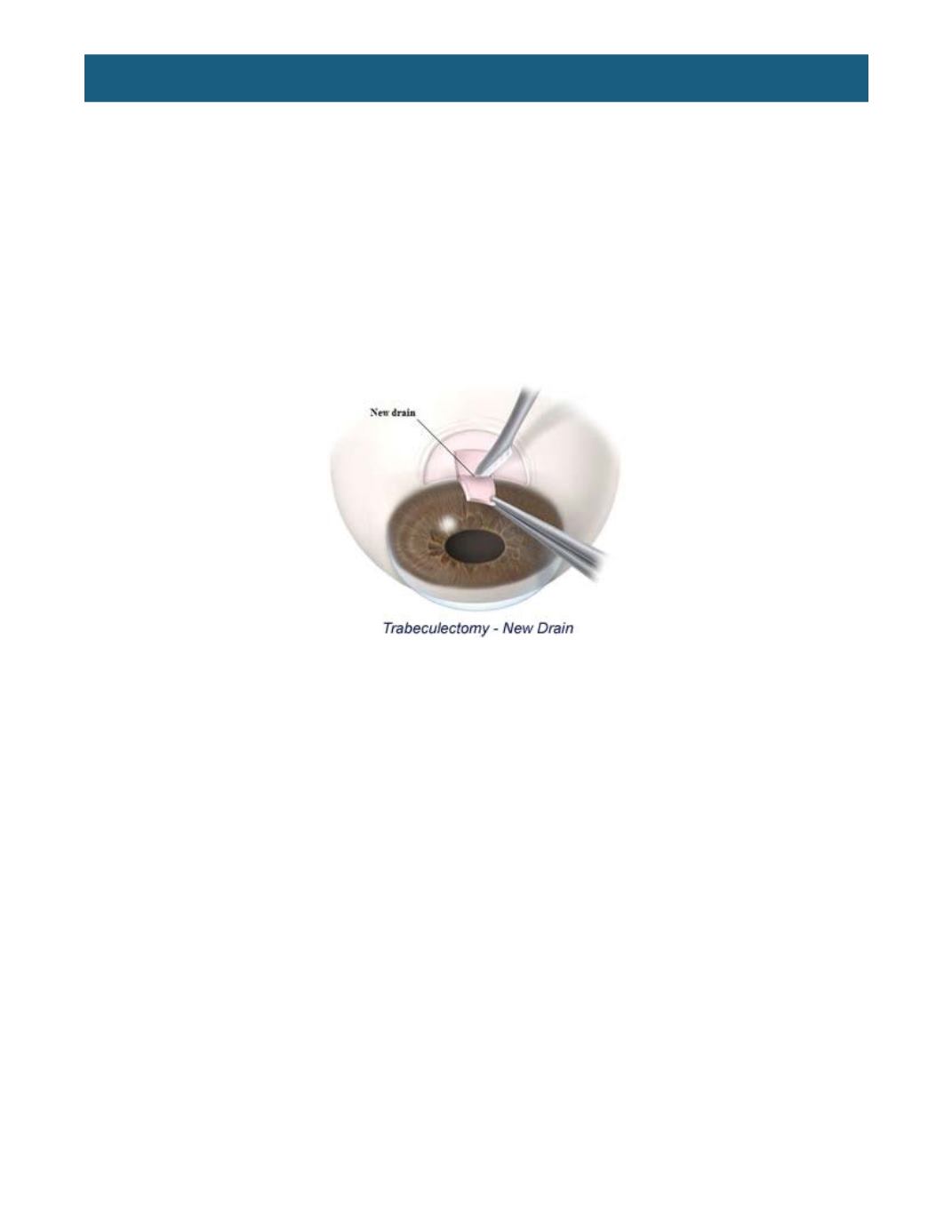
Trabeculectomy:
In a trabeculectomy, the surgeon bypasses the trabecular meshwork and creates a
new drainage system for the eye. A partial-thickness scleral flap is made in the superior part of the eye to
take aqueous from inside the eyes anterior chamber to outside of the eye. This flap, or filter, is then
covered by the conjunctiva, Fluid production within the eye is allowed to continue normally, and pressure
reduction is achieved by allowing the fluid to exit the eye through the new drain, which then creates a
“bleb,” or blister-like area, on top of the sclera where the fluid is drained. Some surgeons apply
medications such as Mitomycin-C or 5-Fluorouracil, or use a collagen implant under the bleb at the time
of surgery. These act to reduce scar formation to allow the bleb to remain open for effective drainage of
aqueous.
Trabeculectomy with Mini Glaucoma Shunt:
This is similar to a trabeculectomy, with the addition of a
metal implant to transport the fluid from the anterior chamber to the area of the bleb instead of a surgical
opening with iridectomy. The advantages and disadvantages to traditional trabeculectomy are under
investigation.
Trabectome:
A relatively new procedure, the trabectome replenishes outflow by unblocking the natural
drainage pathway. A small incision is made across the anterior chamber and this small, needle sized
instrument is placed in position. Diseased tissue is targeted with electrosurgical pulses and the debris is
rinsed away with saline.
Canaloplasty:
Canaloplasty involves making a small incision under a scleral flap. Schlemm’s canal is
identified, then intubated with a cannula, which has a lighted tip or beacon to identify its location as it
passes through the canal. Viscoelastic is passed through 360° to allow for dilation of the canal. A suture
is then often placed with tension inside the canal to improve outflow through the trabecular meshwork.
The scleral incision is closed, reducing the risk of post-operative infection.
Glaucoma implants:
Occasionally, because of abnormal eye anatomy or history of previous eye surgery,
it is not possible for the surgeon to build a new drain from the tissue present within the eye, or in other
circumstances where trabeculectomy was felt to be doomed to fail. Under these circumstances, a plastic
tube may be inserted into the eye to act as a new drain. The tube ends of these devices are inserted into
the eye, and the other end of the implant is secured underneath the conjunctiva. This creates a new route
for the aqueous fluid to bypass the trabecular meshwork and exit the eye. These devices include shunts
(hollow tubes that improve drainage) and valves (which include the ability to “shut off” drainage).
Combined glaucoma and cataract surgery:
Since glaucoma most often occurs in older individuals,
many patients develop cataracts that affect the quality of their vision. Although the correction of vision
loss requires surgery, the good news is that cataract surgery may
successfully reverse the vision loss
associated with the cataract. However, the glaucomatous damage will still be present after cataract