


 13 / 22

© 2016, BSM Consulting
11
Basics of Glaucoma
Generalized enlargement of the cup may be the earliest change detected in glaucoma. This enlargement
can be difficult to appreciate unless previous photographs or diagrams are available. Progressive
photographs of the discs are provided through a variety of laser scans of the optic nerve. However,
particular attention should be given to the neuroretinal rim. Even with a normal sized CD ratio, any
evidence of thinning or notching of the rim, particularly in the vertical directions, could be consistent with
early glaucomatous change. In addition, any disc hemorrhages (splinter or Drance hemorrhages) on the
neuroretinal rim could be an indication of glaucomatous damage. Comparing one eye to the fellow eye is
useful because disc asymmetry is unusual in normal individuals.
The optic nerve head may be evaluated using a variety of diagnostic instruments. The following
discussion describes various methods of such evaluations:
Ophthalmoscopy:
The optic disc can be examined clinically with a direct ophthalmoscope, an indirect
ophthalmoscope, or a slit-lamp biomicroscope using a posterior pole lens.
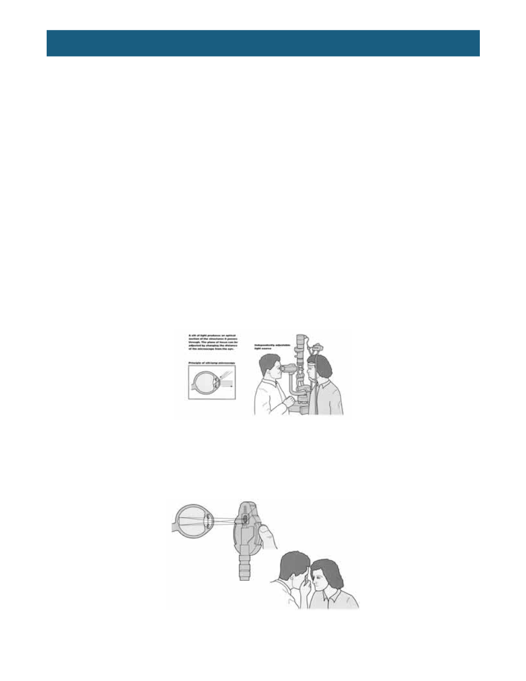
The most preferred method of examination of the optic nerve is the slit lamp combined with lens. A Hruby
lens, a posterior pole contact lens, or a 60-, 66-, 78-, or 90-diopter lens can be used, along with other
handheld lenses. The slit beam, rather than diffuse illumination, is useful for determining subtle changes
in the contour of the nerve head. This system provides high magnification, excellent illumination and a
stereoscopic view of the disc. Slit-lamp techniques require patient cooperation and moderate pupil size
for adequate visibility of the disc.
The direct ophthalmoscope provides a view of the optic disc through a small pupil. In addition, when used
with a red-free filter, it enhances detection of the nerve fiber layer of the posterior pole. However, the
direct ophthalmoscope does not provide sufficient stereoscopic detail to detect subtle changes in optic
disc topography.
The indirect ophthalmoscope is used for examining the optic disc in young children, uncooperative
patients, individuals with high myopia, and individuals with substantial opacities of the media. The indirect
ophthalmoscope can detect cupping of the optic nerve, but, in general, optic nerve cupping and pallor are
less distinct than with slit-lamp methods. The magnification often is inadequate for detecting subtle or
localized details important in the evaluation of glaucoma. Thus, the indirect ophthalmoscope is not
recommended for routine use in examining the optic disc.