


 16 / 22

© 2016, BSM Consulting
14
Basics of Glaucoma
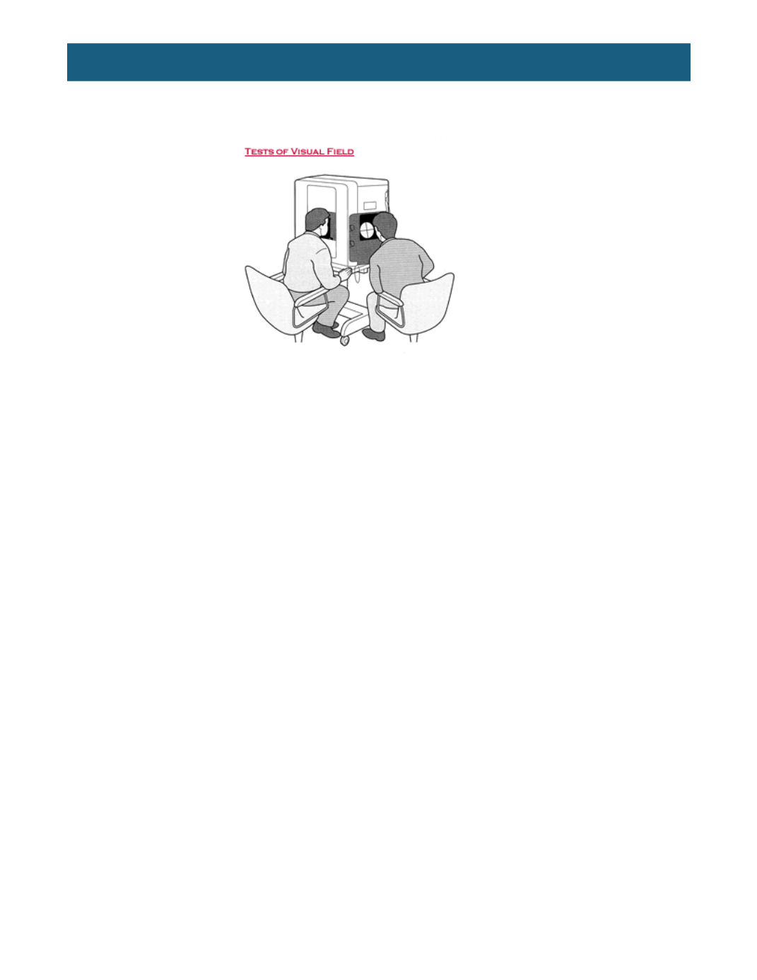
and SITA Fast) help increase the accuracy and decrease the time of the examination. Occasionally, the
size of the test area or the size of the stimulus are changed to achieve a test with more information for a
given patient.
In a reliable visual field, the exam printout will most often reveal a blind spot at 10 to 15 degrees temporal
to the center of fixation. This blind spot is a normal finding and is located at the point where the optic
nerve exits the eye. Other areas of visual field defects are identifiable by the intensity of the grayscale of
the printout: the darker the defect, the more absolute the vision loss. Scotoma is the term for a visual field
defect, indicating partial to complete blindness in the corresponding spot in the island of vision.
Even though the test is fully automated, the technician should be on hand to ensure patient positioning
and comfort, and that the patient is clear on the instructions and follow-through. Reliability of the visual
field test is measured by:
•
Fixation losses:
The number of times the patient indicates having seen the stimulus during testing
of the blind spot (where there should be no vision).
•
Positive errors:
The patient has indicated that the stimulus was seen when none was offered.
•
Negative errors:
The patient does not signal having seen the stimulus in spots previously
identified.
No matter which test format, the job of interpretation falls to the physician. Visual field test results, used in
conjunction with other tests, assist the physician in the diagnosis and treatment of glaucoma.
MEDICAL MANAGEMENT OF GLAUCOMA
The goal of current glaucoma therapy is to preserve visual function by lowering IOP to a level that is
unlikely to produce further damage to the nerve. The treatment regimen that achieves this goal with the
lowest risk, fewest side effects, and least disruption of the patient’s life, is the physician’s most frequent
choice. The “target pressure goal” should actually be a range, with an upper IOP limit that the physician
feels is unlikely to lead to further damage of the optic nerve in a given patient.
The more advanced the glaucomatous process on initial presentation, the lower the target pressure
generally needs to be to prevent further progression. This more aggressive target is to minimize the risk
of progressive glaucomatous damage. Some feel that once the optic nerve is damaged, it is more likely to
incur additional damage. An initial reduction in the IOP of 20-40 percent from baseline is frequently the
goal of the physician; however, reduction of IOP to the target pressure range does not guarantee that
progression will not occur. The target pressure range requires constant reassessment and change, as
IOP fluctuations, optic nerve changes, and visual field progression dictate.