


 10 / 22

© 2016, BSM Consulting
8
Basics of Glaucoma
Applanation measurements are safe, easy to perform, and are relatively accurate in most clinical
situations. The Goldmann applanation tonometer is the most valid and reliable device currently available.
Because applanation does not displace much fluid or substantially increase the eye pressure, this method
is relatively unaffected by ocular rigidity.
Excessive fluorescein results in wide mires and inaccurately high readings, whereas inadequate
fluorescein leads to low readings. When applanating eyes with less than 3 diopters of cylinder power, the
0- and 180-degree marks are placed parallel to the white lines on the prism holder. Marked corneal
astigmatism causes an elliptical fluorescein pattern. To obtain an accurate reading, the clinician should
rotate the prism so the red mark on the prism holder is set at the least curved meridian of the cornea
along the negative axis. Alternately, two pressure readings taken 90 degrees apart can be averaged.
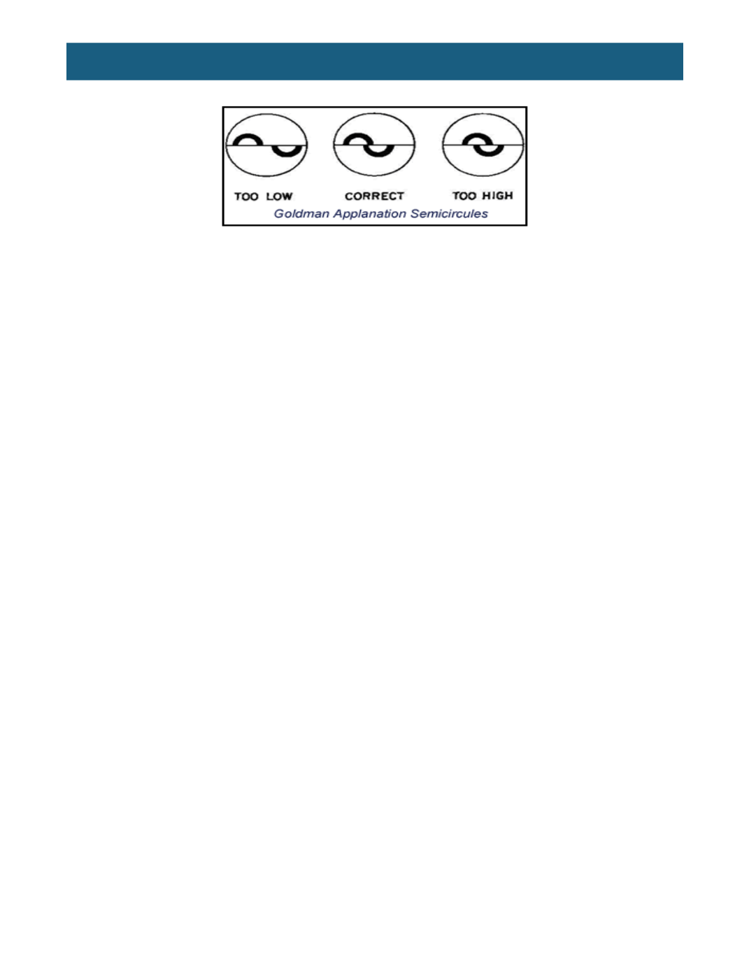
Once the patient and tonometer are prepared, the technician slowly moves the tonometer toward the
central cornea by watching for the reflection of the tonometer in the patient’s pupil. Once the tonometer is
about a quarter-inch away from the central cornea, the technician looks for the ghost mires in the left
ocular.
The accuracy of applanation tonometry is reduced in certain situations. Corneal edema predisposes to
falsely low readings, whereas pressure measurements taken over a corneal scar will be falsely high.
Additionally, tonometry over a soft contact lens gives falsely low values. Alterations in scleral rigidity may
compromise the accuracy of measurements. For example, applanation readings that follow scleral
buckling procedures might be inaccurately low.
Increased central corneal thickness (CCT) may give an artificially high IOP measurement, while
decreased CCT may give an artificially low IOP measurement. IOP measured after photorefractive
keratectomy or laser in situ keratomileusis may be reduced because of changes in the corneal thickness
induced by these procedures. CCT affects the measurements provided by the Goldmann tonometer,
Perkins tonometer, pneumatonometer, non-contact tonometer, and Tonopen.
The portable, counterbalanced Perkins tonometer can be used with the patient either upright or supine. It
is similar to the Goldmann tonometer, using a split-image device and fluorescein staining of the tears.
Hand-held, it is very useful when the patient is unable to reach the slit lamp. Centering the prism tip on
the cornea is often accomplished by first placing the forehead rest above the appropriate brow. The prism
tip may require adjustment after viewing the ghost mires off the central cornea.
Other Methods of Measuring IOP
Non-contact (air-puff) tonometers
measure IOP without touching the eye, measuring the time
necessary for a given force of air to flatten a given area of the cornea. Readings obtained with these
instruments can vary widely, and they often overestimate IOP. Non-contact tonometers are frequently
used in large-scale glaucoma-screening programs or by non-medical health care providers. The
advantages of these tonometers are ease of use and no need for anesthetic drops.
Portable electronic applanation devices,
such as the Tonopen, applanate a very small area of the
cornea and are particularly useful in the presence of corneal scars or edema, or when it is difficult to use
other methods of applanation due to the positioning or location of the patient (e.g., in a hospital bed).
These instruments register a series of pressure readings and average them to display the IOP in