


 7 / 22

© 2016, BSM Consulting
5
Basics of Glaucoma
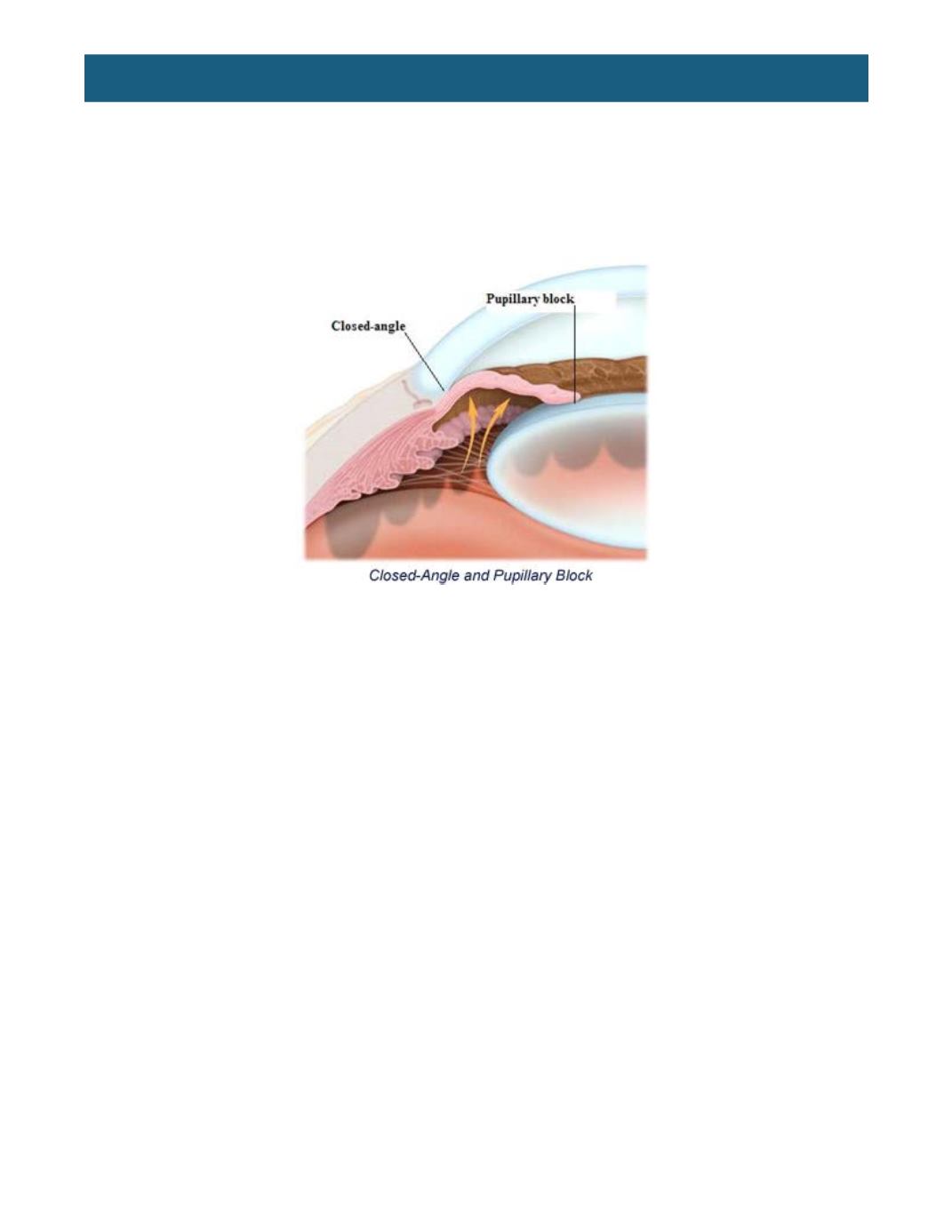
lens in such a way that the aqueous cannot flow through the pupil into the anterior chamber. This is called
pupillary block, which results in the buildup of pressure in the posterior chamber pushing the peripheral
iris forward against the filtration angle. Any fluid in the anterior chamber cannot drain out through the
trabecular meshwork because it is also blocked by the iris. This situation results in sudden, acute
elevation of the ocular pressure called acute angle-closure glaucoma. In patients with narrow angles,
angle-closure glaucoma also can result from natural dilation of the pupil in dim illumination, after dilation
of the pupils in an eye exam, or due to other medications such as decongestants, antidepressants, or
anti-anxiety drugs.
During an attack of angle-closure glaucoma, patients might experience the following symptoms:
•
Red and painful eyes
•
See halos around lights, indicating possible corneal edema
•
Severe headache and nausea
A physician might attempt to break the attack initially by constricting the pupil and lowering the pressure
medically in the office, and then often a laser or incisional surgical procedure.
With regular eye examinations, angle-closure glaucoma is preventable by monitoring the anterior
chamber depth. Once the angles have been deemed “narrow,” the workup technician may have the eye
physician recheck the angles with a slit lamp and gonioscopy prior to dilation. If the angles are
appositional, and therefore putting the patient at risk for an attack of angle closure, a laser peripheral
iridotomy may be performed. In this procedure, the ophthalmologist makes a small hole in the peripheral
iris with the laser, thereby allowing for drainage of the aqueous from behind the iris to in front, where it
can then reach the trabecular meshwork.
It is more common to find narrow angles in hyperopic patients than in those who are nearsighted. The
average axial length of the eye is 23.5 millimeters, and hyperopes tend to have shorter eyes than
emmetropes. Therefore, it is likely that as a cataract grows and pushes the iris forward into the chamber,
the anatomic structures become more crowded, and the outflow of aqueous decreases. Generally, the
risk of angle closure is greater, with more hyperopia.
A rarer type of anatomically narrow-angle glaucoma is plateau iris syndrome. This can be determined
only after a laser iridotomy. If the angle is still narrow after the iridotomy, this could suggest that the iris is
blocking the trabecular meshwork due to the structure of the iris, not to trapped aqueous pushing the iris
forward. Treatments for this disorder are less established but may include laser iridoplasty to reshape the
peripheral iris, pilocarpine drops to constrict the pupil, or cataract surgery to free up space in the front of
the eye.