


 10 / 22

© 2016, BSM Consulting
8
Modern Glaucoma Surgery
Cataract surgery alone is known to lower IOP, so studies evaluating the iStent require a control group of
patients undergoing cataract surgery alone without iStent implantation. In one such study, 72% of stented
patients versus 50% of cataract-only patients had IOP≤21 mmHg on no medications at 12 months, with
66% versus 44%, respectively, achieving IOP reduction ≥ 20%
. 18However, the average number of
medications used by the stent group decreased from 1.6 preoperatively to 0.2 at 12 months. A longer
study found that the proportion of patients achieving IOP≤21 mmHg without medications at 24 months
was 61% in the iStent group and 50% in the cataract-only group—this difference was statistically
significant, but only marginall
y. 19In the same study, the proportion of patients with IOP reduction ≥20% at
24 months was 53% versus 44%, respectively, which was not statistically significant. However, the
average number of medications used by the stent group decreased from 1.6 preoperatively to 0.3 at 24
months.
The procedure of implanting the iStent during cataract surgery is quite safe. Many eyes will have mild,
transient hyphema, and some will have brief hypotony as well. These rarely require intervention and
usually resolve quickly. The postoperative course and follow-up schedule is similar to that of cataract
surgery alone.
Based on these outcomes, this procedure may be best suited for glaucoma patients whose IOP is well
controlled on two or more medications who are undergoing elective cataract surgery and wish to reduce
their glaucoma medical burden, as the procedure lowers IOP only modestly but can significantly reduce
the need for IOP-lowering medications.
OTHER OUTFLOW PROCEDURES
There are several procedures that have been developed but never became widely accepted, or fell out of
favor, mostly due to limited efficacy, unacceptable complication rates, difficulty mastering the procedure,
or a combination of these reasons. They are briefly discussed here.
Deep Sclerectomy ± Viscocanalostomy
This is essentially a modified, blebless trabeculectomy. Once the scleral flap is made, it is extended into
the peripheral cornea and a second deeper scleral flap is fashioned under the first one and then cut
away. This creates an empty pocket in the sclera when the first flap is closed. Before it is closed,
however, a section of the peripheral cornea is thinned down leaving just Descemet’s membrane under the
scleral flap. In the process, the outer wall of Schlemm’s canal is removed under the flap as well, and
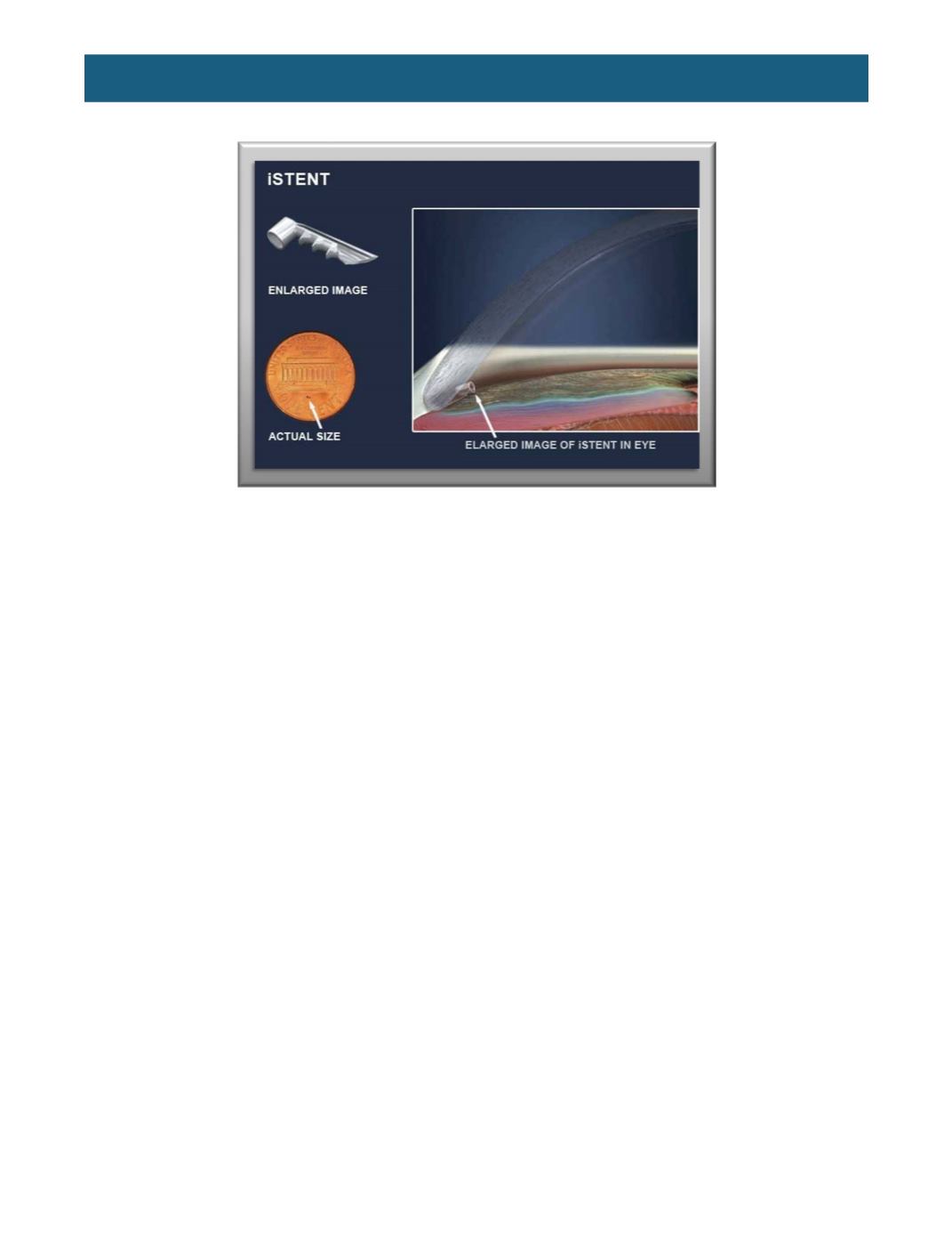
Figure 5.
The iStent device.
(From
http://www.eosc.org/iStent.html)